The Norwood scale: understanding male pattern hair loss
A stage can describe the pattern of hair loss. It cannot, by itself, tell you how quickly it will change or how many grafts you need.

What the Norwood scale measures
The Hamilton–Norwood scale is a way of describing common patterns of male hair loss. It looks at recession around the temples and front, thinning over the crown and how those areas join as loss becomes more extensive. It helps make a discussion or photographic record more consistent.
It is not a diagnosis and it does not capture every pattern. Diffuse thinning, inflammation, sudden shedding and patchy hair loss need an assessment rather than simply a stage number. Our male pattern baldness guide explains the underlying condition.
Norwood stages 1–7
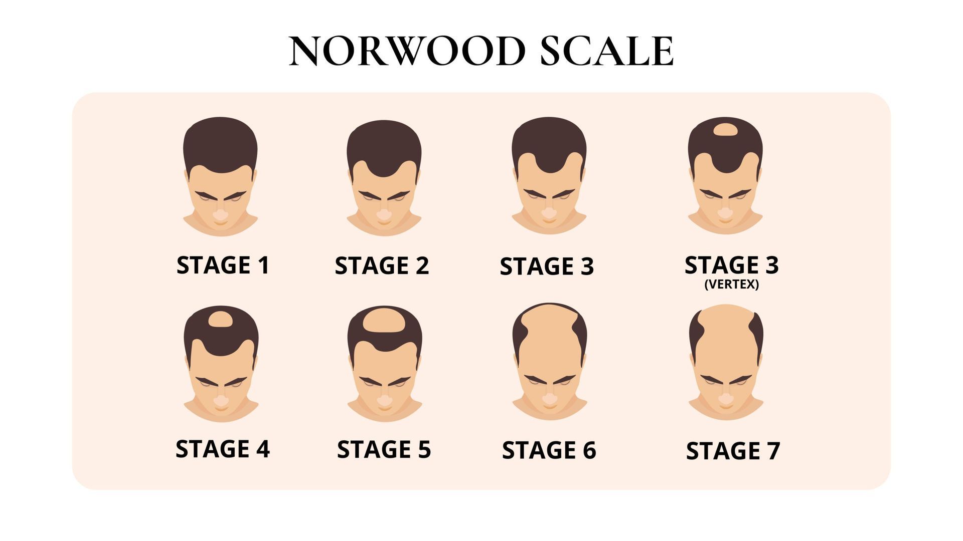
The drawings below show the head from above, with the forehead at the bottom. Dark areas represent hair coverage; lighter areas show recession or loss. They describe patterns, not a timetable. You may stay at one stage, and your own hair loss may not match a drawing exactly.
Stage 1: little or no recession
The hairline is largely intact, with little change at the temples. This is the starting reference in the scale. A photograph from this point can be useful later, but the number alone says nothing about whether your hairline will change.
Stage 2: the corners begin to recede
The change is mainly at the temples. The corners sit a little further back, while the middle of the hairline remains further forward, giving the front a more noticeable M shape. Compare photographs taken under similar conditions before deciding that a small difference is new hair loss.
Stage 3: deeper recession at the temples
The bare or sparsely covered areas at the corners are more pronounced than in stage 2. The centre of the hairline may still project forwards. Looking only at the front can miss what is happening elsewhere, so photographs from above and behind are useful too.
Stage 3 vertex: a separate crown pattern
Stage 3 vertex describes a pattern in which thinning is more noticeable at the crown. Temple recession does not need to extend beyond stage 3. This is a separate pattern in the classification; it is not a compulsory step between stages 3 and 4.
Stage 4: front and crown loss, with hair between
There is more recession at the front and a clearer area of thinning or loss at the crown. A band of hair still crosses the middle of the scalp, separating the two areas. That remaining band is the main feature to look for when comparing this pattern with the later stages.
Stage 5: the separating band becomes narrower
The areas at the front and crown are larger. Hair still separates them, but the band is narrower or less dense than in stage 4. An overhead view helps show this change; a front-facing photograph may give very little information about the crown.
Stage 6: the front and crown areas join
The band that separated the front from the crown has largely disappeared, leaving one connected area of loss across the top. Hair remains at the sides and back. How much of that hair can be used for transplantation needs its own assessment; the stage does not measure donor supply.
Stage 7: a narrower band remains around the scalp
This is the most extensive pattern described by the scale. Hair remains in a narrower horseshoe-shaped band around the sides and back. If surgery is being considered, the discussion needs to focus on achievable coverage and priorities rather than assuming the whole top can regain its original density.
There are also type A patterns, in which the hairline moves backwards without a separate bald area developing at the crown. If your hair is thinning more evenly across the scalp, a single Norwood number may be a poor description.
How to make photographs useful
Take pictures from the front, both temples, above and the crown. Keep the lighting, camera angle, distance and hair length as similar as you can. Wet hair, a fresh haircut or strong overhead light can make the same scalp look very different.
Compare photographs taken months apart, rather than repeatedly checking day to day. Record when shedding or recession first became noticeable and whether there have been changes in health, medication or weight. These details can be more useful at a consultation than a self-assigned stage.
What a Norwood number cannot tell you
The scale does not measure donor density, the thickness of individual hairs or how much the appearance bothers you. It also does not reliably predict the final pattern. A family history is useful context, but it is not a timetable for your own hair loss.
DermNet's explanation of male pattern hair loss describes the inherited tendency and gradual miniaturisation involved. Thinning may coexist with other causes of shedding, so a change in appearance deserves a proper history and examination.
How it helps with treatment planning
Treatment starts with establishing the cause and discussing your priorities. Medical treatment may be considered for pattern hair loss; benefits, adverse effects and the need for continued use should be discussed with an appropriate clinician. Surgery is one possible option, not an automatic consequence of reaching a particular stage.
There is no reliable conversion from a Norwood stage to a graft number. The size of the area, desired hairline, existing hair and available donor supply all matter. Our donor area guide explains why preserving that supply is central to planning.
For more extensive loss, a realistic plan may favour the front and leave lighter coverage elsewhere. For early recession, the question may be whether to monitor or treat the underlying loss before considering surgery. A low hairline chosen now can be difficult to support if loss progresses.

When to arrange an assessment
Arrange an assessment if you are uncertain about the cause, if the pattern is changing or if you want to compare treatment options. The NHS advises speaking to a GP about hair loss before attending a commercial clinic. Sudden patches, scalp symptoms or rapid diffuse shedding should not be assumed to be ordinary male pattern loss.
For a Cardiff consultation, bring your photographs, medication list and details of any previous treatment. Our locations page distinguishes consultation rooms from the sites used for surgery.
Questions and answers
Does stage 2 mean I need a transplant?
No. A mildly receded hairline alone does not establish a need for surgery. Your age, pattern over time, examination and preferences all inform the discussion.
Will everyone reach stage 7?
No. The scale describes patterns of increasing extent; it does not mean that every person passes through all seven stages.
Can the scale be used for women?
It is mainly used for male pattern hair loss. Women commonly have different patterns, and clinicians may use other scales alongside the examination.
Can I diagnose my stage from a selfie?
A selfie may show the front but miss the crown and diffuse thinning. Consistent photographs from several angles can support, but cannot replace, clinical assessment.